Measuring tumor ecosystems in motion.
Cancer cells do not progress, persist, or die in isolation. Their behavior is shaped by tissue architecture, extracellular matrix, vessels, immune context, systemic cues, and local signals that vary across space and time.
The Davies Cancer Lab builds living tissue models, biosensor imaging platforms, and computational frameworks to watch these interactions unfold and identify earlier opportunities to intercept progression or therapeutic escape.
What makes a cell trajectory become a disease trajectory?
We study progression and drug response as dynamic tissue-level processes. A single tumor cell follows a path through a changing physical and biochemical landscape; the goal is to learn which features of that landscape determine fate.
- Trajectory: What path does a tumor cell follow as it grows, invades, survives treatment, or dies?
- Boundaries and guides: Which physical structures, cell contacts, tissue niches, and matrix features steer that path?
- Signals: What signals are present, who produces them, and when do they become decisive?
- Response: How do cancer-cell state, host context, and therapy converge to determine drug response?
We approach these questions by combining biosensors, living tissue models, and computation: biosensors report live pathway state; tissue models preserve tumor-host context; computation turns movies into quantitative models.
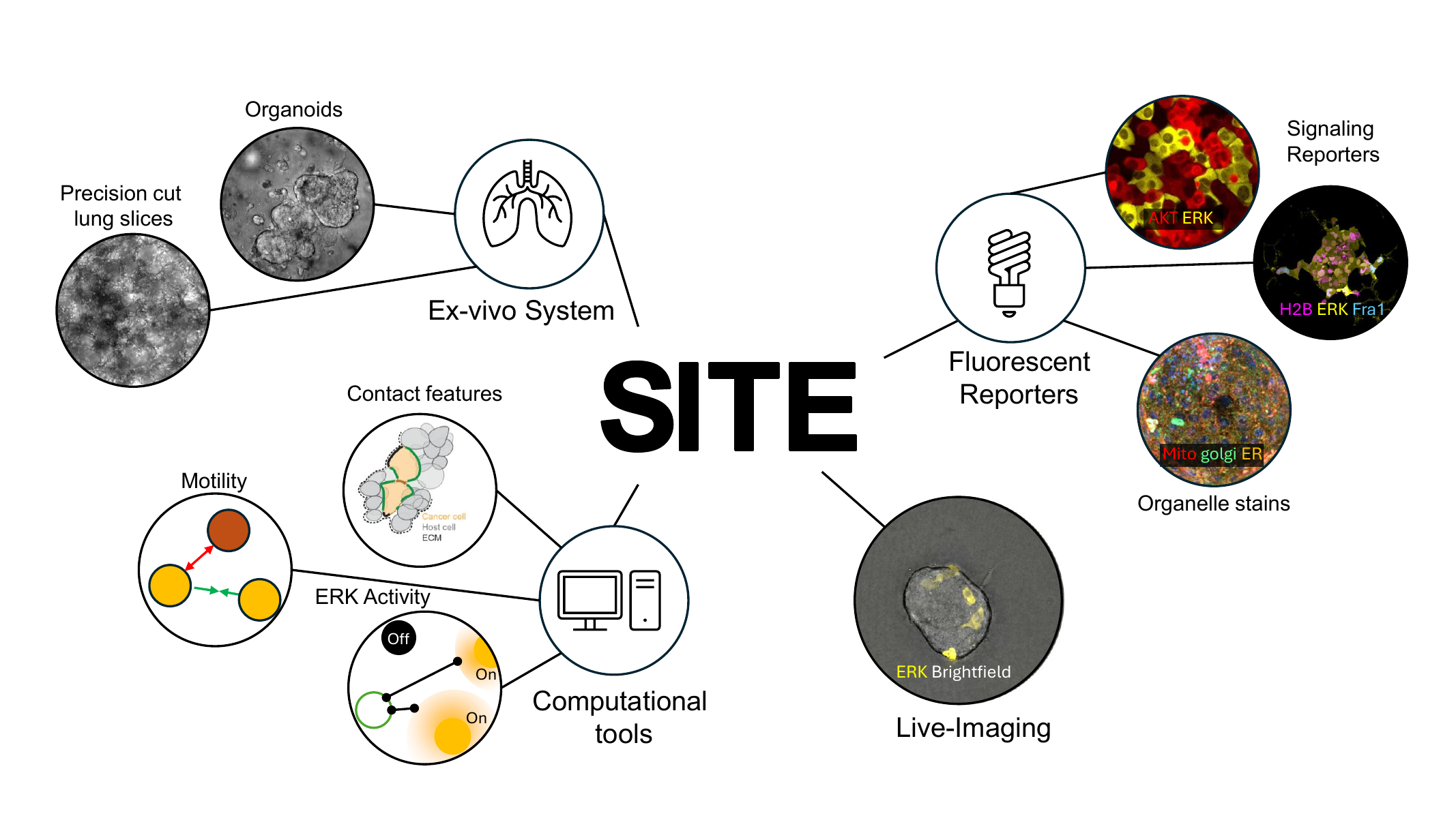
Serial Imaging of Tumor and microEnvironment.
SITE is the lab’s route from living tissue movies to quantitative tumor microenvironment biology. The platform combines ex vivo and tissue-like models, biosensors, time-lapse live-cell imaging, segmentation, tracking, microenvironmental featurization, and data-driven modeling.
From model to measurement
Whole living tissues and tissue-like systems preserve tumor-host context while reporter cells make pathway state directly visible. Instead of measuring only a static endpoint, SITE follows individual cells through their local histories: where they are, who they contact, how they signal, and whether they survive, die, or adapt.
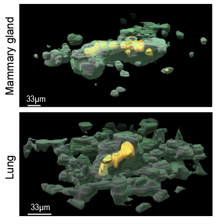
- Living models: Tumor reporter cells are introduced into mammary, lung, and related tissue contexts for longitudinal imaging.
- 4D measurement: Cell segmentation, tissue segmentation, tracking, and reporter quantification convert movies into single-cell time series.
- Interaction analysis: Cancer-host contact, cancer-cancer contact, microenvironmental proximity, signaling, and fate are measured together.
LungSITE and tissue-scale analysis
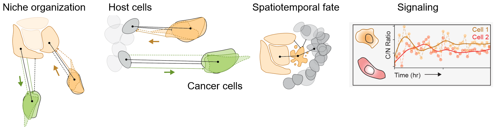
LungSITE extends SITE to cancer cells growing in living lung tissue. Custom analysis routines resolve tumor reporter cells, host tissue, 3D position, local niche, signaling state, and fate across time.
What gets measured?
- Position and niche: Tumor-host contact, tumor-tumor contact, and tissue proximity.
- Cell signaling: Biosensor-derived dynamics such as ERK and AKT activity.
- Spatiotemporal fate: Death, persistence, motility, and adaptation in local context.
- Drug response: How local histories reshape survival after perturbation.
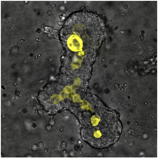
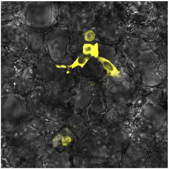
Watching metastatic response in the lung microenvironment.
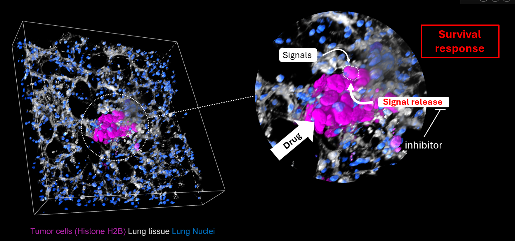
Osteosarcoma lung metastasis remains a major clinical challenge. The lab uses LungSITE to study how metastatic tumor cells respond to host-derived signals, local tissue architecture, and therapy in a living lung environment.
Why lung metastasis?
Metastatic osteosarcoma survival remains poor, and standard treatment has changed slowly. This creates a strong need for models that can reveal how tumor-host interactions regulate survival, death, and resistance in the metastatic lung niche.
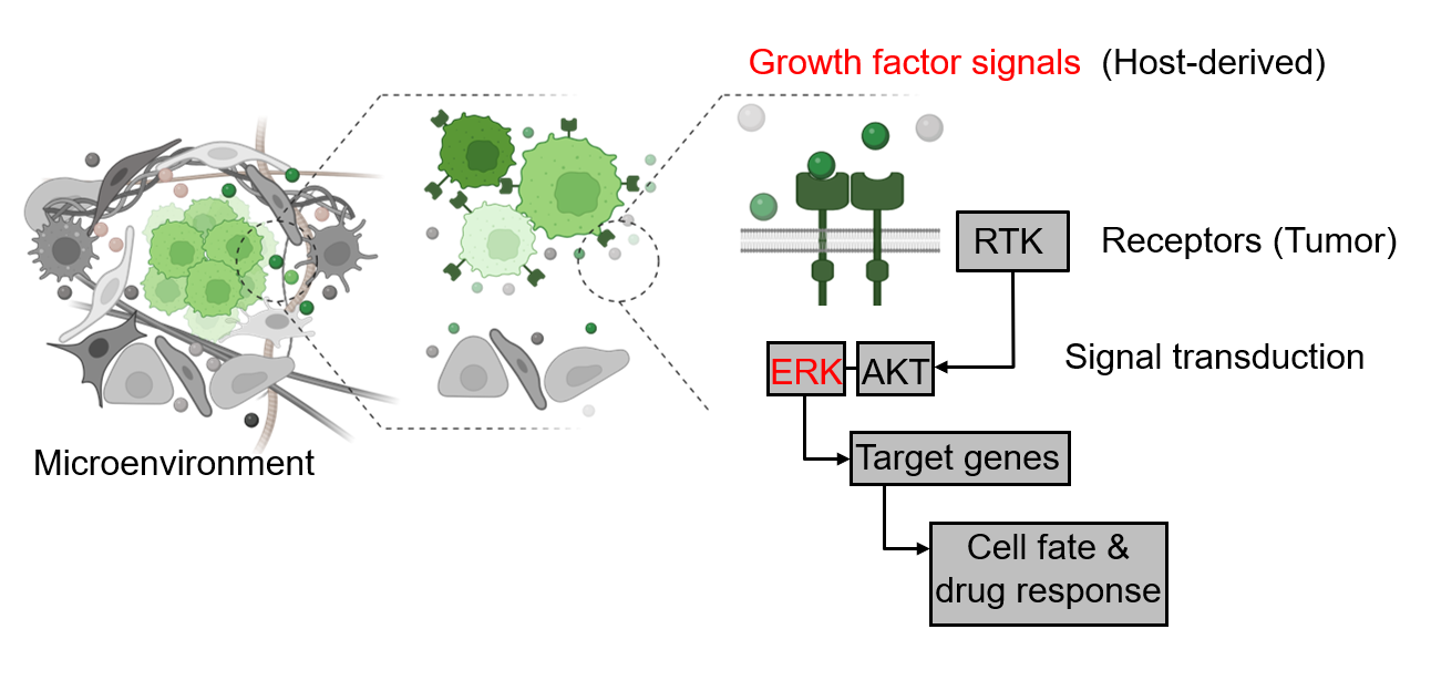
- Host-derived growth-factor signaling can alter tumor-cell pathway activity.
- RTK, ERK, and AKT signaling provide measurable routes linking microenvironmental cues to cell fate.
- Longitudinal imaging makes it possible to distinguish immediate drug killing from adaptive survival.
Drug response is dynamic.
One of the central lessons from LungSITE is that therapy response is not only an endpoint. Tumor cells can die, persist, change position, alter signaling, and experience new signals released by damaged tissue or dying cells. Those time-dependent changes can create survival states that are invisible in static assays.
- Measure before and after treatment: Track the same lesion over time rather than comparing unrelated snapshots.
- Connect signaling to fate: Ask whether ERK/AKT dynamics predict death or persistence.
- Test rational combinations: Use response dynamics to identify when upstream signaling blockade may improve tumor killing.
Mechanism and intervention
The current working model is that treatment can reshape the lung tumor microenvironment: drug-induced death and tissue damage release signals that feed back through receptor signaling, activate ERK/AKT, and support persistence in a subset of tumor cells. This points to a therapeutic strategy: pair direct tumor-cell killing with interventions that block the adaptive signaling response that follows.
Computational details, software, and model explainers live at CancerDynamics.org.
The formal lab site introduces the biological questions and experimental platforms. CancerDynamics.org hosts deeper materials: SITE resources, trajectory modeling, MMIST, active tissue models, public repositories, and publication-linked resources.